
Surgery as a Management Option for Post-Chikungunya Chronic Synovitis Presenting as a Dorsal Wrist Mass

https://doi.org/10.65989/698120vyvbkx
Abstract
Introduction: Chikungunya fever is a mosquito-borne viral illness that commonly causes acute febrile disease followed by persistent musculoskeletal manifestations. Localised mass-like synovial lesions of the wrist following Chikungunya infection are rarely reported.
Case presentation: We report a 40-year-old female who developed a progressively enlarging dorsal wrist mass following serologically confirmed Chikungunya infection. Despite conservative treatment, she developed significant functional limitations. Imaging demonstrated inflammatory synovitis and extensor tenosynovitis. Surgical excision was performed, and histopathology confirmed chronic inflammatory synovitis.
Discussion: Post-Chikungunya chronic synovitis is thought to result from persistent immune-mediated inflammation. While medical management remains first-line, surgery may be indicated in selected cases with Localised disease and functional impairment.
Conclusion: This case highlights surgery as a valuable management option for Localised post-Chikungunya chronic synovitis presenting as a dorsal wrist mass.
Article
- Introduction
Chikungunya virus infection is Characterised by acute fever, polyarthralgia, and myalgia. Although most patients recover completely, a substantial proportion develop chronic inflammatory musculoskeletal manifestations such as arthritis, tenosynovitis, and synovitis. These chronic sequelae can persist for months or years and significantly affect quality of life. Localised mass-forming synovitis of the hand and wrist following Chikungunya infection is uncommon and may pose a diagnostic challenge. We present a rare case of post-Chikungunya chronic synovitis presenting as a dorsal wrist mass requiring surgical management.
- Case Presentation
A 40-year-old female presented with a progressively enlarging lump over the dorsal aspect of her right hand and wrist. She had a documented history of Chikungunya fever, confirmed on 1st June by ELISA IgM positivity. The acute illness was Characterised by one week of high-grade fever followed by post-viral arthralgia and myalgia involving both upper and lower limb joints.
A few weeks after the Febrile episode, she noticed a Localised swelling over the dorsum of the right hand, which gradually increased in size. There was no history of trauma, local infection, or previous similar swellings. The swelling was associated with pain and significant functional limitation, particularly during finger extension, most notably involving the middle finger, despite treatment with non-steroidal anti-inflammatory drugs, corticosteroids, and other conservative measures.
Clinical examination revealed a soft to firm, non-fluctuant mass measuring approximately 5.4 × 3 cm over the dorsal aspect of the right wrist and hand. The overlying skin was normal with no erythema or warmth. Wrist movements were mildly restricted due to pain, and no neurovascular deficits were detected.
Ultrasonography of the right hand demonstrated moderate subcutaneous edema over the dorsal aspect with mild fluid accumulation surrounding the extensor tendons, particularly involving the middle finger. Increased synovial vascularity was noted, consistent with active inflammatory synovitis and mild extensor tenosynovitis. No Localised collections, abscesses, or discrete mass lesions were identified.
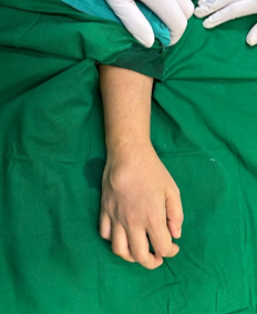
Figure 1: Pre-Operative appearance of the lump
Given the progressive symptoms and functional impairment, surgical exploration and excision were planned. Under general Anaesthesia and with the use of a pneumatic tourniquet to achieve an avascular field, a longitudinal dorsal incision was made. Careful dissection revealed a firm, capsulated mass with proximal extension beneath the extensor retinaculum up to the distal forearm, incorporating the extensor tendons distally. Complete excision was performed with meticulous dissection, and the specimen was sent for histopathological examination. The wound was closed using absorbable monofilament subcuticular sutures.
Histopathological examination revealed synovial tissue with hyperplasia, dense chronic inflammatory infiltrate, vascular proliferation, and mild perivascular chronic inflammation with lymphoid follicle formation. There was no evidence of suppuration, granuloma formation, myxoid degeneration, or neoplasia. These findings supported a diagnosis of chronic inflammatory synovitis.
Based on clinical history, imaging findings, and histopathology, a diagnosis of post-Chikungunya chronic synovitis of the right wrist was established.
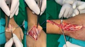
Figure 2: Intraoperative appearance of the lump
Discussion
Chikungunya virus infection is well recognized for its chronic rheumatological sequelae, with persistent arthralgia, arthritis, tenosynovitis, and synovitis occurring in up to 30–60% of patients, particularly middle-aged women. The underlying pathogenesis is believed to involve viral persistence and immune-mediated inflammatory responses within synovial tissues.
Histopathological features such as synovial hyperplasia, lymphocytic infiltration, vascular proliferation, and lymphoid follicle formation have been described in post-Chikungunya chronic synovitis and closely resemble inflammatory arthropathies such as rheumatoid arthritis. Localised mass-like presentations are rare and may mimic ganglion cysts, neoplastic lesions, or infective processes.
While medical management remains the cornerstone of treatment, surgical intervention is indicated in selected cases with diagnostic uncertainty, failure of conservative therapy, or significant functional impairment due to Localised synovial hypertrophy or tendon involvement. In the present case, progressive restriction of finger extension and extensive extensor tendon involvement necessitated surgical excision, which provided both diagnostic confirmation and symptomatic relief.
Conclusion
Post-Chikungunya chronic synovitis can rarely present as a Localised dorsal wrist mass causing significant functional limitation. In such cases, surgical excision serves as both a diagnostic and therapeutic modality. Clinicians in endemic regions should consider post-Chikungunya inflammatory pathology in the differential diagnosis of wrist and hand masses.
Patient Consent
Written informed consent was obtained from the patient for publication of this case report and accompanying images.
Ethical Approval
Formal ethical approval was not required for this case report in accordance with institutional policy.
Funding
No funding was received for this study.
Conflict of Interest
The authors declare no conflicts of interest
References
- Simon F, Javelle E, Cabie A, et al. Chikungunya infection: an emerging rheumatism among travelers returned from endemic areas. Rheumatology (Oxford). 2011;50(10):1756–1762.
- Rodríguez-Morales AJ, Cardona-Ospina JA, Urbano-Garzón SF, Hurtado-Zapata JS. Prevalence of post-Chikungunya chronic inflammatory rheumatism: a systematic review and meta-analysis. Arthritis Care Res (Hoboken). 2016;68(12):1849–1858.
- Hoarau JJ, Jaffar Bandjee MC, Krejbich Trotot P, et al. Persistent chronic inflammation and infection by Chikungunya arthritogenic alphavirus in spite of a robust host immune response. J Immunol.2010;184(10):5914–5927.
- Labadie K, Larcher T, Joubert C, et al. Chikungunya disease in nonhuman primates involves long-term viral persistence in macrophages. J Clin Invest. 2010;120(3):894–906.
- Bouquillard E, Combe B. A report of 21 cases of rheumatoid arthritis following Chikungunya fever. A prospective study. Ann Rheum Dis. 2009;68(9):1505–1507.
- Ganu MA, Ganu AS. Post-chikungunya chronic inflammatory arthritis—our experience with DMARDs over two years. J Assoc Physicians India. 2011;59:83–86.
- Elsinga J, Gerstenbluth I, van der Ploeg S, et al. Long-term Chikungunya sequelae in Curaçao: burden, determinants, and a novel classification tool. J Infect Dis. 2017;216(5):573–581.