
Intraneural Venous Malformation of the Ulnar Nerve
https://doi.org/10.65989/455708dnofwq
Abstract
Background
Intraneural venous malformations affecting peripheral nerves particularly the ulnar nerve is exceedingly rare and may present as compressive neuropathies. Proper clinical evaluation, diagnostic imaging and histopathology play key roles, and microsurgical resection is mainstay of treatment.
Case presentation
A 20-year-old right-hand dominant female presented with right forearm cramps and intermittent numbness radiating to the hand for 3 months duration. 3 years back she has undergone surgical exploration of same region following imaging studies due to similar symptoms suspecting a vascular lesion . Previous surgical exploration has identified a clinically suspected venous malformation within and deep to the flexor tendons, for which debulking and decompression were performed and histology was confirmed as an arteriovenous malformation. After a three-year symptom free interval, similar but worsening symptoms gradually recurred .Ultrasound scan with doppler followed by MRI showed lesion recurrence of venous malformation abutting the ulnar neurovascular bundle. Re-exploration revealed an extensive intraneural venous malformation involving the ulnar nerve. Microsurgical debulking and decompression were performed, with complete excision confirmed on histology. The patient recovered well, with improved sensation and normal hand function.
Conclusion
Intraneural venous malformations of the ulnar nerve, though rare, should be considered in the differential diagnosis of atypical neuropathic symptoms. Ultrasound with Doppler and MRI facilitates diagnosis and surgical planning, and microsurgical decompression with nerve preservation offers favorable outcomes.
Article
Introduction
Venous malformations are congenital low-flow vascular anomalies that predominantly involve skin and soft tissues. Their intraneural occurrence, especially within peripheral nerves like the ulnar nerve, is extremely rare and diagnostically challenging. Only a limited number of cases have been reported in the literature. These lesions can mimic common entrapment neuropathies, leading to delays in diagnosis and treatment. Doppler ultrasound and MRI (Magnetic Resonance Imaging) serves as a critical imaging modality, while histopathology is definitive. Surgical decompression through microsurgical techniques remains the mainstay of management. This case report highlights the diagnostic and therapeutic journey of a recurrent intraneural venous malformation involving the ulnar nerve in a young patient.
Case Presentation
A 20 year old right hand dominant schoolgirl, with no significant medical or allergy history, has been initially evaluated in year 2022 with clinical symptoms of right forearm cramps during writing, intermittent radiating forearm pain and numbness. There was no history known trauma or vascular symptoms. Examination revealed a soft, sub-centimeter, reducible lump on the volar aspect of the right wrist. The motor and sensory functions and distal vascularity were intact.
Following clinical evaluation imaging studies were done. Ultrasound scan with Doppler was suggestive of well-defined heterogeneously hypo echoic lesion (15 × 8 mm) deep to flexor tendons with internal vascularity. MRI Revealed abnormal tortuous vascular channels encircling the flexor digitorum superficialis (FDS) and profundus (FDP) tendons of right distal forearm. DSA (Digital Subtraction Angiography) was attempted but no abnormalities noted. Nerve conduction study (NCS) was also normal.
Exploration identified a venous malformation located within and beneath the FDS and FDP muscles. Debulking and carpal tunnel decompression were performed in year 2022. Histopathology confirmed an arteriovenous malformation. The patient remained symptoms free for three years.
In year 2025 march, she sought medical attention due to re-experiencing similar symptoms for 3 months duration with worsening symptoms comparing to the past experience. Repeat nerve conduction study was also normal. Ultrasound with doppler was suggestive of residual or recurrence of previous malformation. MRI showed recurrent lesion of venous malformation in the flexor compartment of right forearm abutting the ulnar neurovascular bundle.
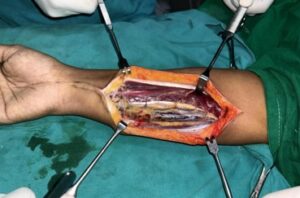
Fig – 1 (Intraoperative finding of intraneural venous malformation)
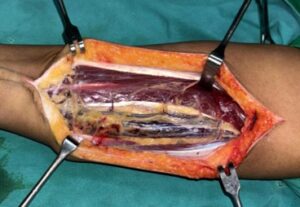
Fig – 2 (Close look)
Re-exploration was performed through a longitudinal incision along the ventromedial forearm, incorporating the previous scar in line with the ulnar nerve. Under magnification, the neurovascular lesion was carefully exposed, and an epineurotomy was performed to access the intraneural venous malformation. The lesion was meticulously dissected with controlled debulking while preserving neural integrity. Hemostasis was secured, and the specimen was sent for histopathological evaluation, which confirmed a venous malformation with complete excision.
The postoperative course was uneventful and sensory symptoms improved, and she was subsequently followed up with occupational therapy and physiotherapy.
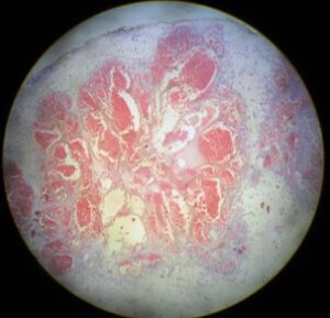
Fig – 3 (Histological Appearance of the excised venous malformation)
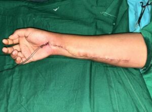
Fig – 4 (after skin closure)
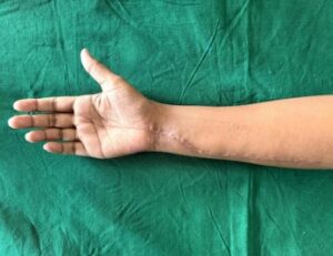
Fig – 5 (At the follow up visit)
Discussion
Venous malformation (VM) is the most common type of congenital vascular malformation (CVM) with an incidence of 1 to 2 in 10,000 and a prevalence of 1% [1]
Although present at birth, they are not always clinically evident until later in life and tend to grow in concert with the child and without spontaneous regression [2]
Venous malformations arising from the peripheral nerve are a rare type of vascular malformation [3] and this can cause compression neuropathies.
Venous malformation involving median nerve causing acute carpal tunnel syndrome has been reported [4]
Compression of the ulnar nerve is most commonly caused by ganglions, lipomas, anomalous tendon and muscles, trauma related to an occupation and arthritis in the upper limb; however, nerve compression in the upper limb and, more importantly, in the forearm by a vascular lesion is rare [5]
Doppler ultrasound (US) and magnetic resonance imaging (MRI) are key imaging methods used to characterize and diagnose venous malformations [6]
Duplex Ultrasound is a useful, non-invasive imaging technique and should be used as the first modality when investigating the presence of a vascular malformation, especially for superficial lesions or those in the extremities [7]
Magnetic resonance angiography (MRA) is useful in mapping the venous system and has supplanted the diagnostic role of conventional angiography in many cases [8] Angiography remains an adjunct imaging modality and important for cases requiring therapeutic intervention of venous malformations [6]
Treatment options include endoscopy assisted or open microsurgery, sclerotherapy and ablative techniques [6]
Conclusion
This case underscores the rare and challenging nature of intraneural venous malformations affecting the ulnar nerve. MRI is invaluable in diagnosis and surgical planning, while histology confirms the nature of the lesion. Microsurgical nerve-preserving decompression provides favorable outcomes. Vigilant long-term follow-up is essential due to high recurrence potential.
The patient reported satisfaction with the surgical outcome, particularly noting improvement in sensation. She appreciated the multidisciplinary follow-up and occupational therapy support, which helped her return to daily activities.
Declarations
None
ORCID
Kanil weranja https://orcid.org/0009-0003-9431-7454
Gayan Ekanayake https://orcid.org/0000-0001-8420-7073
Uditha Wickramadewa https://orcid.org/0009-0002-5878-7881
Eshani Dabare https://orcid.org/0009-0000-2256-8893
Ethics approval and consent to participate
Not applicable
Consent for publication
Informed written consent for publication and accompanying images was obtained from the patients prior to collecting information.
Availability of data and material
All data generated analyzed during this study are included in this published article
Competing interests
The authors received no financial support for the research, authorship, and/or publication of this article.
References
- Vikkula M, Boon LM, Mulliken JB. Molecular genetics of vascular malformations. Matrix Biol 2001;20:327-35. 10.1016/S0945-053X(01)00150-0 [DOI] [PubMed] [Google Scholar]
- Richter GT, Friedman AB. Hemangiomas and vascular malformations: current theory and management. Int J Pediatr 2012;2012:645678. [DOI] [PMC free article] [PubMed]
- González Porto SA, González Rodríguez A, Midón Míguez J. Intraneural Venous Malformations of the Median Nerve. Arch Plast Surg. 2016 Jul;43(4):371-3. doi: 10.5999/aps.2016.43.4.371. Epub 2016 Jul 20. PMID: 27462571; PMCID: PMC4959981.
https://pmc.ncbi.nlm.nih.gov/articles/PMC4959981/
- Hariri A, Cohen G, Masmejean EH. Venous malformation involving median nerve causing acute carpal tunnel syndrome. J Hand Surg Eur Vol. 2011 Jun;36(5):431-2. doi: 10.1177/1753193411405946. Epub 2011 Apr 13. PMID: 21490029.
https://pubmed.ncbi.nlm.nih.gov/21490029/
DOI: 10.1177/1753193411405946
- Saquib M, Khan N, Mir A. Ulnar Nerve Compression in the Proximal Forearm By An Intraneural Arteriovenous Malformation. Plastic Surgery Case Studies. 2015;1(2):43-44. doi:10.1177/2513826X1500100207
- Behravesh S, Yakes W, Gupta N, Naidu S, Chong BW, Khademhosseini A, Oklu R. Venous malformations: clinical diagnosis and treatment. Cardiovasc Diagn Ther. 2016 Dec;6(6):557-569. doi: 10.21037/cdt.2016.11.10. PMID: 28123976; PMCID: PMC5220204.
https://pmc.ncbi.nlm.nih.gov/articles/PMC5220204/
doi: 10.21037/cdt.2016.11.10
- Lee BB, Baumgartner I, Berlien P, et al. Diagnosis and Treatment of Venous Malformations. Consensus Document of the International Union of Phlebology (IUP): updated 2013. Int Angiol 2015;34:97-149
[PubMed] [Google Scholar]
- Herborn CU, Goyen M, Lauenstein TC, et al. Comprehensive time-resolved MRI of peripheral vascular malformations. AJR Am J Roentgenol 2003;181:729-35. 10.2214/ajr.181.3.1810729 [DOI] [PubMed] [Google Scholar]