Staged Excision of an Intraosseous Hemangioma of the Maxilla via Gingival and Intranasal Approaches

https://doi.org/10.65989/243840dksawg
Abstract
A 15-year-old female presented with a right-sided nasal osseous lesion, managed initially via a right upper gingival excision. Histopathology confirmed an intraosseous hemangioma. Clinical follow-up identified a remaining portion of the lesion, necessitating a planned second stage. Two years later, a piecemeal excision of the remnant was performed using an intranasal approach, safely preserving the infraorbital nerve. The patient experienced expected right-sided facial swelling post-operatively, underscoring the need for routine post-operative care and monitoring following maxillary surgery.
Article
Introduction
Intraosseous hemangiomas are relatively uncommon, slow-growing, benign vascular tumors. While they typically arise in the vertebral column or calvarium, primary intraosseous hemangiomas of the facial skeleton, particularly the maxillary bone, are exceedingly rare. Management primarily involves surgical excision, with the surgical approach dictated by the tumor’s size, location, and proximity to vital structures. Complete resection is necessary to prevent persistent growth; however, staged interventions may be required to safely address remnant tissues. This report details the surgical management and follow-up of a right maxillary intraosseous hemangioma in an adolescent female, treated via a staged surgical protocol.
Case Presentation
A 15-year-old female patient presented in April 2024 with an osseous lesion located on the right side of the nose. Initial clinical and radiological evaluation led to a surgical plan for the excision of the right nasal osseous lesion under general anesthesia.
The first surgical intervention was performed on October 17, 2024.A right upper gingival incision was made to access the surgical site. A vascularized bony lesion over the right maxilla was identified, excised, and sent for histopathology. Hemostasis was successfully achieved, and the gingiva was closed using 5-0 Monocryl sutures.
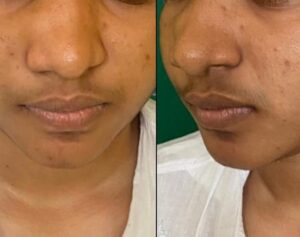
Figure 1: Pre-operative clinical appearance. Multi-panel view demonstrating the right-sided nasal osseous lesion prior to surgical intervention.
During an interval follow-up on May 16, 2025, clinical assessment identified a remaining part of the lesion, leading to a planned second-stage excision. On May 21, 2026, at the age of 17, the patient underwent a piecemeal excision of the remaining right maxillary osseous lesion.An intranasal approach was utilized to access the right maxilla, ensuring the infraorbital nerve and associated vessels were directly visualized and safeguarded. An intraoral incision was also utilized and closed with 4/0 Monocryl, followed by the insertion of a nasal pack to maintain hemostasis.
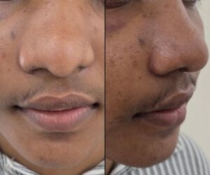
Figure 2: Post-operative assessment at two weeks
Results
Histopathological analysis of the decalcified bony tissue fragments from the primary surgery revealed lamellated bony fragments containing a vascular lesion. The lobules consisted of thin-walled blood vessels lined by flat endothelial cells without any malignant features, yielding a definitive conclusion of an intraosseous hemangioma.
Following the primary surgery in 2024, the patient was discharged with a three-week follow-up plan. She was prescribed oral Co-amoxiclav 625mg TDS. The duration of this postoperative prophylactic antibiotic therapy was closely monitored to ensure strict compliance with antimicrobial stewardship guidelines, effectively preventing infection while strictly limiting unnecessary prolonged antibiotic exposure.
Following the second-stage surgery in May 2026, the recovery plan involved a 12-day at-home convalescence. During the one-week review on May 29, 2026, the patient presented with anticipated right-sided facial swelling, which was successfully managed conservatively.
Discussion
Surgical management of maxillary intraosseous hemangiomas requires careful planning to balance complete tumor removal with the preservation of facial aesthetics and function. The initial gingival approach allowed direct access but necessitated a subsequent intervention for the remnant lesion. The staged intranasal approach utilized for the remaining tissue proved highly beneficial, allowing for safe piecemeal excision without external facial scarring, while affording direct protection of the infraorbital nerve.
The postoperative development of right-sided facial swelling is an expected sequela of maxillary surgery. Vigilant postoperative monitoring remains important in maxillofacial procedures to manage localized edema and ensure the continued functional integrity of the safeguarded infraorbital nerve.
Learning Points:
- Intraosseous hemangiomas of the maxilla are rare but must be considered in the differential diagnosis of vascularized bony facial lesions.
- A staged excision is a safe and viable strategy when remnant tissue is identified, prioritizing anatomical preservation over overly aggressive single-stage resections.
- The intranasal approach provides an excellent, scarless route for addressing right maxillary lesions while allowing for the safe safeguarding of the infraorbital nerve.
- Postoperative monitoring is essential to manage localized regional swelling and ensure the functional integrity of preserved neurovascular structures.
The complete eradication of maxillary intraosseous hemangiomas can be safely achieved through a staged, multi-approach surgical strategy. Utilizing an intranasal approach for remnant lesions offers a highly effective pathway that minimizes aesthetic morbidity and protects vital neurovascular structures. Close postoperative observation remains essential for managing expected regional swelling and ensuring uncomplicated wound healing.
Declarations
None
ORCID
Gayan Ekanayake https://orcid.org/0000-0001-8420-7073
G.R.B.S.Wijewardhana https://orcid.org/0009-0005-0562-1409
K.D.B Meegolla https://orcid.org/0009-0002-0061-5681
Yulugxan https://orcid.org/0000-0001-5119-3293
Ethics approval and consent to participate
Approval from the institutional review board/ethics committee was obtained for the surgical management and follow-up detailed in this report. The patient and their legal guardians provided informed consent to participate.
Consent for publication
Written informed consent was obtained from the patient and her legal guardians for the publication of this case report and any accompanying images or data.
Availability of data and material
All data generated or analyzed during this study are included in this published article.
Competing interests
The authors declare that they have no competing interests.
Funding
No funding was received for the preparation or publication of this case report.
References
- Kim, T.-G., Whangbo, C.-H., Ye, M.-K., & Shin, S.-H. (2026). Management Considerations in Nasal Bone Intraosseous Cavernous Hemangioma: A Case Report and Literature Review. Case Reports in Medicine, 2026. https://doi.org/10.1155/carm/1774878
- Hamamin, O. S., Abdulrahman, S. W., Aziz, J. A. A., Rashid, M. J., & Hashan, M. (2025). Cavernous hemangioma: a rare case report in the maxillary sinus. Annals of Medicine and Surgery, 88, 809 – 813. https://doi.org/10.1097/ms9.0000000000004302
- Cerqueira, É., Correia, S., Ramalhosa, F., Lopes, T., & Abreu, J. M. (2024). Challenges and Management of Capillary Intraosseous Hemangioma in the Mandibular Symphysis: A Case Report. Cureus, 16. https://doi.org/10.7759/cureus.58035
- Douami, A., Marsafi, O., Belgadir, H., Merzem, A., Nashi, L., Amriss, O., Moussali, N., & Elbenna, N. (2024). Maxillary intraosseous hemangioma: case report. The Pan African Medical Journal, 48. https://doi.org/10.11604/pamj.2024.48.135.43355
- Francois, H. A., Oumaima, B., Rajaa, E. A., Ghita, E. A. H., Bouchra, D., & Malik, B. (2024). INTRAOSSEOUS CAVERNOUS HEMANGIOMA OF THE ZYGOMA: A CASE REPORT. International Journal of Advanced Research. https://doi.org/10.21474/ijar01/18677
- Kim, K.-H., Min, J.-H., Lee, E.-S., Kim, D.-H., & Lim, H.-K. (2024). Intraosseous Hemangioma of the Maxilla, Possibly Misdiagnosed as a Periapical Cyst : A Case Report. The Journal of The Korean Dental Association. https://doi.org/10.22974/jkda.2024.62.11.003
- Li, S. C., Zhang, Y., Liu, H., & Wu, Y. (2024). Primary Intraosseous Hemangiomas of the Maxillary Bone: A Case Report and Literature Review. Ear, nose, & throat journal, 1455613241272466. https://doi.org/10.1177/01455613241272466