
Chronic Chromoblastomycosis of the Upper Limb: Serial Surgical Excision with Split-Thickness Skin Grafting in a 13-Year Refractory Case

https://doi.org/10.65989/961510utqylm
Abstract
Background: Chromoblastomycosis remains one of the more frustrating chronic fungal infections in tropical surgical practice. Although prolonged itraconazole is standard first-line therapy, a subset of patients with extensive, multifocal disease simply do not respond to medical treatment alone — these are the patients who reach the plastic surgery clinic.
Case Presentation: We describe a 65-year-old Sri Lankan male with a 13-year history of chromoblastomycosis involving the left upper limb, referred after failure of prolonged itraconazole therapy and cryotherapy. Over 18 months he underwent four staged procedures comprising wide local excision of multiple lesions with split-thickness skin grafting (STSG). Initial graft take was 95%. Histopathology confirmed the diagnosis, with characteristic sclerotic bodies and pseudocarcinomatous hyperplasia; all specimens were clear of malignancy.
Conclusion: For extensive chromoblastomycosis refractory to medical therapy, serial surgical excision with STSG offers reliable reconstruction with good functional outcomes. The keys are adequate excision margins, careful patient selection, and acceptance that these cases usually require staged procedures rather than a single operation.
Article
- Introduction
Chromoblastomycosis is not a condition most plastic surgeons encounter regularly, but in tropical practice it lands on our doorstep with increasing frequency. It is a chronic granulomatous fungal infection caused by dematiaceous fungi, most commonly Fonsecaea pedrosoi, predominantly affecting agricultural workers [1][2], and is now recognised by the WHO as a neglected tropical disease [3].
The typical presentation is warty, hyperkeratotic plaques or cauliflower-like nodules, usually on the lower limbs, though upper limb involvement is well documented. The hallmark histological finding is the muriform cell, or “copper penny” — a thick-walled, brown, septate structure that is pathognomonic [2][4]. Diagnosis is straightforward once biopsy is performed; management is anything but. First-line itraconazole 200–400 mg daily for 6–24 months is standard, yet cure rates are disappointing — between 15% and 80% depending on the series [5] — and recurrence after apparently successful treatment exceeds 40% [6]. Terbinafine is an alternative, and combination therapy has been advocated for refractory cases, but drug access and long-term compliance remain significant barriers [2].
What concerns the surgeon most is the long-term sequelae: lymphedema, chronic ulceration, secondary infection, and dense fibrosis [4]. More worryingly, disease beyond 10 years carries a documented risk of malignant transformation to squamous cell carcinoma in 2–13% of cases [7][8]. We present this case to illustrate the reconstructive surgical approach to extensive, multifocal disease that has failed conservative management.
- Case Presentation
2.1 Background
Mr. W, a 65-year-old shopkeeper from Mathugama, presented to our Plastic Surgery Unit at the National Hospital of Sri Lanka in late 2024. He had carried a diagnosis of chromoblastomycosis since 2013, initially managed at Colombo South Teaching Hospital with cryotherapy and oral itraconazole. Despite years of treatment — taken intermittently, unsurprising given the duration required — the disease had relentlessly progressed across his left upper limb: the elbow flexor region, volar forearm, dorsum of the hand, and thenar eminence. A 2020 biopsy had confirmed chromoblastomycosis with pseudocarcinomatous hyperplasia and sclerotic bodies within multinucleated giant cells.
His comorbidities included hypertension, type 2 diabetes on metformin, and dyslipidaemia, along with Grade III–IV fatty liver with mild hepatomegaly on abdominal ultrasound and mildly deranged liver enzymes (AST 35 U/L, ALT 53 U/L, bilirubin 1.7 mg/dL) attributed to prolonged itraconazole exposure — a significant consideration when weighing further high-dose antifungal therapy and prolonged postoperative antibiotic cover.
2.2 Clinical Findings
On general examination he was thin but well, clinically euthyroid, with stable vital signs and no pallor or jaundice. The left upper limb showed firm, non-tender, hyperkeratotic plaques with verrucous surfaces and surrounding lichenification. The “black dots” of extruded sclerotic bodies were visible to the naked eye — almost diagnostic in an endemic area [9]. Crucially, there was no regional lymphadenopathy, no distal neurovascular compromise, and hand function was preserved: our goal was not only to clear disease but to maintain a functional limb.
Fresh biopsies from three sites (elbow, thenar eminence, dorsum of hand) all confirmed chromoblastomycosis with pseudoepitheliomatous hyperplasia, chronic granulomatous inflammation, and numerous muriform sclerotic bodies, with no dysplasia or malignancy —reassuring, though with a 13-year history we remained vigilant.
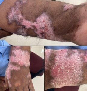
Figure 1- Preoperative appearance of the left upper limb
2.3 Surgical Management
The decision to operate was straightforward: the disease was extensive, multifocal, had failed years of medical therapy, and the risk of malignant transformation in a 13-year lesion was a genuine concern. We planned staged wide local excision with STSG, accepting from the outset that multiple procedures would be needed. Over 18 months, the patient underwent four procedures:
First Procedure — 20 October 2024. The largest lesion, on the left forearm, was excised under combined supraclavicular block and spinal anaesthesia with a 5 mm margin of clinically normal tissue, and resurfaced with a meshed STSG from the left thigh applied without quilting. With meticulous haemostasis, limb elevation, and 48 hours of IV co-amoxiclav, day-2 inspection showed ~95% take. The donor site healed within three weeks and he was discharged on day 6 with oral antibiotics and standard analgesia.
Second Procedure — March 2025. Additional left forearm lesions were excised and grafted (left thigh STSG, supraclavicular block). Day-5 discolouration and blistering over the dorsum of the hand was managed conservatively with curettage, drainage, and Vaseline dressings; the graft settled and healed.
Third Procedure — 8 November 2025. Disease had progressed to the left arm, forearm, and dorsum of the hand. All affected lesions were excised and resurfaced with STSG from the right postero-lateral thigh, protected in a backslab, with good take on inspection.
Fourth Procedure — 14 June 2026. Four lesions over the left elbow flexor region and thenar eminence were excised under general anaesthesia. Left thigh STSG resurfaced the elbow and thenar defects; the dorsum of the hand was closed primarily with 3-0 nylon. He was discharged on day 2 after an uneventful recovery.
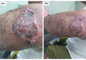
Figure 2- The left elbow region during postoperative wound care following the fourth procedure, showing the healing grafted bed with marginal epithelialization and healthy granulation (a, b).
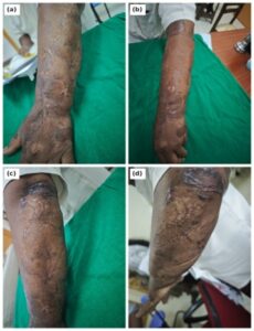
Figure 3- Postoperative appearance of the left upper limb at follow-up: matured split-thickness skin grafts over the forearm and dorsum of the hand (a, b) and the elbow region (c, d), with no contracture and no clinical evidence of recurrent disease
2.4 Postoperative Course and Follow-up
All excised specimens confirmed chromoblastomycosis with clear margins and no evidence of malignant transformation. At the most recent follow-up the grafts had matured well, colour match was acceptable, there was no contracture limiting movement, and no clinical evidence of residual or recurrent infection (Figure 2). He has been counselled on long-term surveillance: with recurrence rates above 40% [6], these patients must be followed for years, not months.
- Discussion
3.1 When to Stop Medical Therapy and Operate
There is no hard rule for when to abandon medical therapy for surgery. The decision rests on disease extent, failure of a reasonable antifungal trial (at least 6–12 months of compliant treatment), patient preference, and complications such as chronic ulceration, secondary infection, or concern for malignancy. Here, his hepatic steatosis and abnormal bilirubin made continued high-dose itraconazole unattractive. Posaconazole has been reported in refractory cases [10], but availability and cost remain barriers in our setting.
3.2 Surgical Technique and the Malignancy Question
Wide local excision with at least a 5 mm margin of healthy-appearing tissue is essential: chromoblastomycosis extends beyond the visible lesion, and incomplete excision is the commonest cause of post-surgical recurrence [6]. All specimens should be sent for histology, both to confirm the diagnosis and to verify clear margins.
We chose STSG over full-thickness grafts or local flaps for several reasons: the defects were too large for primary closure; STSG survives better on a potentially suboptimal bed; meshing allows fluid egress and reduces seroma risk; and the thigh donor site heals reliably and can be re-harvested — as demonstrated over 18 months here.
Any patient with chromoblastomycosis beyond 10 years should be considered at risk for squamous cell carcinoma. Azevedo and colleagues reported SCC in seven patients, all with disease duration exceeding a decade [7]; the mechanism is thought to be chronic inflammation-mediated DNA damage, and coexisting pseudocarcinomatous hyperplasia can make the histological distinction challenging [8]. Every specimen from our patient was scrutinised for dysplasia — none was found, but the risk persists for any future recurrence.
3.3 Graft Survival and Practical Lessons
STSG failure in contaminated wounds is well documented; postoperative infection is the strongest independent risk factor for graft failure (odds ratio 48.3) [11], with Gram-negative colonisation by Pseudomonas or Klebsiella particularly problematic [12][13]. Here, complete excision of diseased tissue, meticulous haemostasis, limb elevation, and co-amoxiclav prophylaxis in a well-nourished, immunocompetent patient worked in our favour.
Three practical points deserve emphasis. First, accept that extensive chromoblastomycosis usually requires staged procedures — clearing everything in one sitting risks prolonged anaesthesia, donor-site morbidity, and compromised graft outcomes. Second, not every defect needs a graft: the small recurrent elbow lesion we managed with simple elliptical excision and primary closure healed uneventfully. Third, compliance with follow-up is as important as compliance with medication — regular review for at least 2–3 years is needed to detect recurrence early.
- Conclusion
Extensive chromoblastomycosis refractory to medical therapy remains a challenging problem in tropical surgical practice. This case demonstrates that a planned programme of serial wide local excision with split-thickness skin grafting can achieve good functional and cosmetic outcomes even in long-standing multifocal disease. The keys to success are adequate excision margins, meticulous technique, appropriate patient selection, and acceptance that multiple procedures may be necessary. Given the risk of malignant transformation in long-standing lesions, early surgical referral should be considered for patients with extensive or refractory disease.
Informed Consent: Written informed consent was obtained from the patient for publication of this case report and accompanying images.
Conflicts of Interest: None declared.
Funding: None.
References
- Queiroz-Telles F, de Hoog S, Santos DW, et al. Chromoblastomycosis. Clin Microbiol Rev. 2015;28(3):586-611. doi:10.1128/CMR.00032-16
- Garcia Garcia SC, Rincon SG, Fernandez Martinez R, Cifuentes Luna C. Chromoblastomycosis. In: StatPearls [Internet]. Treasure Island (FL): StatPearls Publishing; 2024 Mar.
- World Health Organization. Chromoblastomycosis: Key Facts. WHO Fact Sheets. 15 November 2023. Available from: https://www.who.int/news-room/fact-sheets/detail/chromoblastomycosis
- Queiroz-Telles F, Esterre P, Perez-Blanco M, et al. Chromoblastomycosis: an overview of clinical manifestations, diagnosis and treatment. Med Mycol. 2009;47(s1):3-15. doi:10.1080/13693780802538001
- Centers for Disease Control and Prevention. Clinical Overview of Chromoblastomycosis. CDC Fungal Diseases. 30 January 2026. Available from: https://www.cdc.gov/chromoblastomycosis/hcp/clinical-overview/
- Wang TJ, Pica EM, Salzer JS, et al. A case of recurrent chromoblastomycosis treated with multiple surgical management options. Dermatopathology. 2026;13(1):10. doi:10.3390/dermatopathology13010010
- Azevedo CM, Marques SG, dos Santos PAL, et al. Squamous cell carcinoma derived from chronic chromoblastomycosis in Brazil. Clin Infect Dis. 2015;60(10):1500-1504. doi:10.1093/cid/civ104
- Rosenbach M, Kovarik C. Chromoblastomycosis leading to squamous cell carcinoma: An overlooked outcome of a neglected tropical disease. Cutis. 2026;117(5):143-145. doi:10.12788/cutis.0139
- Bonifaz A, Carrasco-Gerard E, Saul A. Chromoblastomycosis: clinical and mycologic experience of 51 cases. Mycoses. 2001;44(1-2):1-7.