
Management of a Brachial Plexus Neurofibroma by Piecemeal Resection

https://doi.org/10.65989/588439dbajmc
Abstract
Brachial plexus tumors are rare and may arise from intrinsic neural elements or adjacent compressive structures. Neurofibromas, benign peripheral nerve sheath tumors, often involve nerve fascicles, making excision challenging with risk of neurological deficit. We report a 46-year-old female with a 5-year history of a progressively enlarging right supraclavicular mass and ulnar sensory impairment. Imaging revealed a well-defined mass related to subclavian vessels. Surgical exploration identified origin from the C8 root with C7 tethering. Piecemeal excision preserved neural structures. Histology confirmed neurofibroma (S100, NSE positive). Sensory improvement was noted at 3 months. Careful planning is essential for optimal outcomes.
Article
Introduction
Tumors involving the Brachial plexus are uncommon and may arise from either intrinsic neural elements or from extrinsic lesions compressing the plexus. Neurogenic tumors in this region are frequently associated with Neurofibromatosis type 1 (NF-1). Neurofibromas are benign, heterogeneous tumors originating from the connective tissue of peripheral nerve sheaths[1]. In contrast to Schwannoma, which typically displaces adjacent neural structures, neurofibromas tend to involve the nerve fascicles intimately, making surgical excision more challenging and increasing the risk of neurological deficit[2].
The role of pre-therapeutic biopsy in suspected benign nerve sheath tumors remains limited. The diagnostic sensitivity is moderate, and the procedure carries potential risks, including damage to intact nerve fascicles and hemorrhage [2]. Accurate pre-operative differentiation between schwannoma and neurofibroma is therefore crucial, as it significantly influences surgical planning and the expected functional outcome following tumor excision [3].
We report a rare case of a solitary neurofibroma arising from the brachial plexus in the absence of NF-1, which was managed with surgical resection.
Case presentation
We present the case of a 46-year-old female with a 5-year history of a right supraclavicular lump associated with sensory impairment in the right upper limb, predominantly along the ulnar nerve distribution, without any motor deficit. Both the size of the lump and the associated symptoms had gradually progressed over time. There was no history of trauma and no constitutional symptoms suggestive of tuberculosis or lymphoma.
Clinical examination revealed a well-defined, firm supraclavicular mass with reduced mobility. Sensory impairment was noted in the ulnar nerve distribution, without evidence of intrinsic muscle atrophy of the hand. Clinically no evidence of peripheral stigmata of Neurofibromatosis. Routine blood investigations were within normal limits, and chest radiography showed no apical lesion
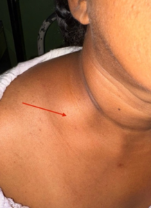
Fig1- Right side Supraclavicular region, well defined solid lump (red arrow)
spindle-shaped mass in the right supraclavicular region measuring 3.0 × 3.7 × 4.8 cm. The lesion was closely related to the subclavian vessels without evidence of significant compression or mass effect on adjacent structures.
The patient was planned for surgical excision under general anesthesia. A supraclavicular incision was made, and the lesion was identified intraoperatively. The tumor was found to arise from the C8 nerve root and was tethered to the C7 nerve root. The accessory nerve and supraclavicular nerve bundle were carefully identified and preserved. Due to the complex anatomical location of the tumor, excision was performed in a piecemeal manner, leaving a small residual portion of the tumor in situ.
Postoperatively, the patient developed a hematoma, which was managed conservatively. She was subsequently discharged with a physiotherapy plan. At the 3-month follow-up assessment, the wound had healed well, and improvement in sensory symptoms was noted.
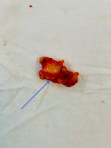
Fig 2- Piece meal excision specimen of brachial plexus neurofibroma(blue arrow)
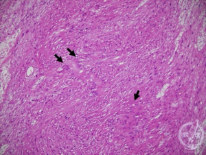
Fig 3- Histology showed a spindle cell lesion composed of long fascicles with focal whorling of cells
Histopathological examination showed a spindle cell lesion composed of long fascicles with focal whorling of cells. The tumor cells exhibited elongated, pointed, wavy nuclei and moderate eosinophilic cytoplasm. Immunohistochemical staining demonstrated diffuse positivity for S100 and NSE. SMA and Desmin were negative in the spindle cells. The Ki-67 proliferative index was approximately 2–3%. No cellular atypia, increased mitotic activity, or necrosis was identified.
Discussion
Peripheral nerve tumors constitute a heterogeneous group of predominantly benign neoplasms that are uncommon in the general population. They are broadly classified into two categories: peripheral nerve sheath tumors and non–nerve sheath tumors. Neurofibromas are characterized by a complex histological composition that includes Schwann cells, perineurial-like cells, and fibroblasts, interspersed with nerve fibers, wire-like collagen bundles, and a myxoid extracellular matrix7). The majority of neurofibromas are solitary lesions (up to 90%) and occur without an association with Neurofibromatosis type. Overall, neurofibromas account for approximately 5% of all benign soft-tissue tumors. [1,5]
Imaging modalities such as magnetic resonance (MR), computed tomography (CT), and positron emission tomography are valuable in the evaluation of peripheral nerve tumors. On CT imaging, schwannomas usually appear hypodense on non-contrast scans and moderately hyperdense following contrast administration, whereas neurofibromas are generally iso- to hypodense on both non-contrast and contrast-enhanced CT images [2]. Most primary tumors involving the brachial plexus require surgical management. Given that neurological deficits may worsen following surgical exploration, operative planning should be carefully guided by the presumed diagnosis and performed by an experienced surgeon. [2,5,6]
Benign tumors of the brachial plexus can be effectively managed with microsurgical techniques, with nerve grafting performed when necessary. In contrast, for malignant lesions, many authors advocate surgical resection with maximal preservation of brachial plexus function, followed by adjuvant radiotherapy and chemotherapy when indicated [3,4].
In the present case, the patient presented predominantly with sensory symptoms without any associated motor deficit. Preoperative assessment and meticulous surgical planning are particularly critical due to the complex anatomical location of the brachial plexus. Preservation of the surrounding neurovascular structures, the extent of dissection, and the degree of residual tumor following resection significantly influence the overall outcome. Importantly, in such cases, patient satisfaction is often determined more by the improvement of clinical symptoms than by the extent of surgical resection alone.
References
1.The Nerve [Internet]. www.thenerve.net. Available from: https://www.thenerve.net/m/journal/view.php?number=190
2.Jarmundowicz, W., Jabłoński, P., & Załuski, R. (2002). Nowotwory splotu ramiennego–taktyka postepowania neurochirurgicznego [Brachial plexus tumors–neurosurgical treatment]. Neurologia i neurochirurgia polska, 36(5), 925–935.
3.Floyd WE. Lesions of the brachial plexus. Journal of the American Society for Surgery of the Hand [Internet]. 2004 May 10;4(2):117–20. Available from: https://www.sciencedirect.com/science/article/abs/pii/S1531091404000488
4.AWu, X., Zhang, Y., Liu, X., Zhang, Q., Yang, Q., Zhang, M., & Lu, P. (2025). Five cases report and literature review of malignant peripheral nerve sheath tumor. Discover oncology, 16(1), 2335. https://doi.org/10.1007/s12672-025-03949-3.
5.Wang, H., Wang, D., Jia, L., Wang, M., Zhang, X., & Shu, P. (2022). Cutaneous malignant peripheral nerve sheath tumor – A case report and literature review. Interdisciplinary Neurosurgery, 28, 101492. https://doi.org/10.1016/j.inat.2022.101492,
6.Grübel, N., Antoniadis, G., Uerschels, A.-K., Gembruch, O., Marschal, V., Deininger, S., König, R., Pala, A., Bremer, J., Dengler, N. F., Reuter, M., Wirtz, C. R., & Pedro, M. T. (2024). Collection of Rare Peripheral Nerve Tumors—Insights from the German Registry. Cancers, 16(14), 2599. https://doi.org/10.3390/cancers16142599