Hibernoma Masquerading as a Conventional Lipoma: A Rare Diagnosis Unveiled by Histopathology

https://doi.org/10.65989/059496lcnpsh
Article
Introduction
A hibernoma is a rare benign tumor composed of brown fatty tissue. It primarily affects young individuals and typically occurs in areas where brown fat is retained after fetal development. The thigh is the most commonly affected area, followed by the trunk, upper extremities, head and neck[1].Some publications detail the imaging findings associated with hibernomas, however, it is not always possible to definitively diagnose a hibernoma, based on imaging alone[2].This case highlights the importance of considering hibernoma in atypical soft tissue masses and the role of surgical excision for diagnosis and treatment.
Case Presentation
A 19-year-old male presented to our clinic with a hemispherical lump at the interscapular region that had persisted for over three years. Apart from the cosmetic concern, he reported no symptoms or functional limitations. Clinical examination revealed a conventional lipoma measuring 4 cm * 1 cm located on the back of the chest, 2 cm below and 6 cm medial to the inferior angle of the left scapula. An ultrasound scan of the lump showed a well-defined, oval, heterogeneous, hyperechoic mass located deep to the subcutaneous fat and superficial to the back muscles, measuring 4.2 cm * 1 cm, without increased vascularity or deep extensions. Since the imaging findings supported a diagnosis of a simple lipoma, we proceeded with lipoma excision under local anesthesia without requesting further imaging investigations. Intraoperatively, the excised lump did not exhibit the typical features of a conventional lipoma, as it was a brownish fatty mass with a well-defined capsule.
Grossly, the tumor had a yellowish lobular appearance. Microscopy revealed a lipomatous tumor composed predominantly of brown fat cells admixed with mature white fat cells. The brown fat cells contained round central nuclei and a multi vacuolated eosinophilic granular cytoplasm. A rich capillary network was present. Nuclear atypia, mitotic activity or lipoblasts were not seen. All these findings conclude that the lump is compatible with a Herbinoma. (Figure 1)
Discussion
A hibernoma is an uncommon, soft tissue tumor that arises from remnants of brown fat, It typically presents as a benign, lobulated, and nontender lesion[1]. Although hibernomas can manifest at any age, they tend to appear most commonly in individuals during their third and fourth decades of life, with male predominance[3]. These tumors typically develop in several areas of the body: the lower limbs (33%), the trunk (23%), the upper limbs (22%), the head and neck (13%), and the abdomen/retroperitoneum (9%) [1].

Figure 1- Hibernoma composed predominantly of brown fat cells. A rich capillary network is seen. (H&E stain x100)
Imaging findings of hibernoma in the lower extremities have been reported numerous times. Ultrasound (US) often reveals a well-defined hyperechoic mass relative to subcutaneous fat. Routine radiography has shown a small soft tissue lesion or swelling without calcification or bone erosion. Doppler imaging has indicated hypervascularity and a hyperechoic mass on ultrasound [2,4]. Complete excision is the definitive treatment for hibernomas, and there have been no reports of local recurrence or aggressive behavior, even in cases of partial excision[1].
Conclusion
This case highlights a rare instance of hibernoma, which was initially misidentified as a conventional lipoma based on clinical and imaging findings. The diagnosis was ultimately confirmed through histopathological examination, highlighting the importance of tissue analysis when intraoperative features differ from expected presentations. Although hibernomas are uncommon, they are benign lesions, and complete surgical excision is a curative treatment.
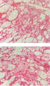
Figure 2,3 : Brown fat cell composed of round central nuclei and a multivacuolated cytoplasm. (H&E stain x400)
Declarations
None
ORCID
Gayan Ekanayake https://orcid.org/0000-0001-8420-7073
Hasod Abeyasinghe https://orcid.org/0009-0005-0159-2831
Consent for publication
Informed written consent for publication and accompanying images was obtained from the patients prior to collecting information.
Availability of data and material
All data generated or analyzed during this study are included in this published article.
Competing interests
The authors declare that they have no competing interests.
Funding
The authors received no financial support for the research, authorship, and/or publication of this article.
References
References
- Furlong MA, Fanburg–Smith JC, Miettinen M. The morphologic spectrum of hibernoma: a clinicopathologic study of 170 cases. The American journal of surgical pathology. 2001;25(6):809-14.
- Yim AC, Sheikh A, Dharmadhikari R, Gravel D, Rakhra K, Gina Di Primio M, et al. Hibernoma: a case series with multimodality imaging and pathologic correlation. Bulletin of the NYU Hospital for Joint Diseases. 2012;70(4):283.
- Park JY, Yi SY, Lee JY, Kwon TJ. A case report of axillary hibernoma: US, CT, MR, and histopathologic findings. Journal of the Korean Society of Radiology (Taehan Yŏngsang Ŭihakhoe chi). 2021;83(2):439.
- AY B, İT K. A rare tumor of the fatty tissue in the axilla; hibernoma. Surgical Chronicles. 2019;24(1).